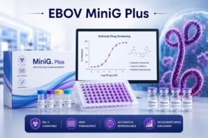
New Ebola Screening Platform, EBOV MiniG Plus, Advances High-Throughput Antiviral Drug Discovery
Ebola virus and challenges of screening antiviral drug
Ebola virus, a member of the Filoviridae family, causes severe viral hemorrhagic disease and requires specialized high-containment laboratories for research involving infectious virus. The development of effective antiviral drugs against high-risk pathogens remains one of the most challenging areas of biomedical research.
Traditional Ebola virus research involving live infectious virus requires biosafety level 4 facilities. These laboratories require specialized infrastructure, strict containment procedures, and highly trained personnel. Although these measures are essential for safety, they also create barriers to large-scale antiviral screening and limit the speed, accessibility, and cost-effectiveness of antiviral drug discovery.
To overcome this limitation, researchers at the Institute of Preventive Medicine, National Defense Medical Center, Taipei, Taiwan developed an enhanced Ebola virus minigenome system, called EBOV MiniG Plus, that improves the detection of viral replication and transcription activity under biosafety level 2 conditions. The platform may provide a more efficient and practical tool for screening potential antiviral compounds without requiring experiments with infectious Ebola virus.
Why Is an Ebola Minigenome System Important?
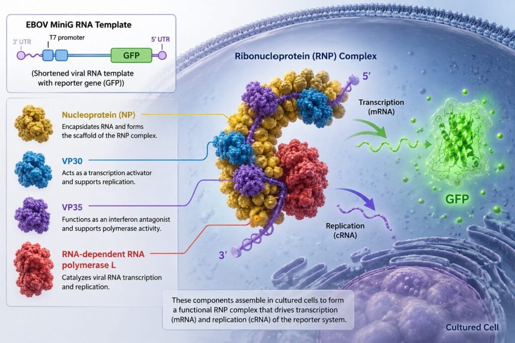
The conventional EBOV MiniG system contains a shortened viral RNA template carrying a reporter gene, such as green fluorescent protein (GFP), together with four essential Ebola virus proteins:
- A nucleoprotein
- VP30
- VP35
- RNA-dependent RNA polymerase L.
So, instead of using the complete infectious virus, the system reproduces essential components involved in Ebola virus transcription and replication.
When these components are introduced into cultured cells, they form a functional ribonucleoprotein complex capable of driving the transcription and replication of the reporter system.
Unlike luciferase-based assays, GFP gene incorporated in the system, can also be detected directly using fluorescence microscopy or flow cytometry without requiring additional substrates or extraction procedures. This may reduce operational complexity and support automated imaging-based screening approaches.
GFP expression therefore serves as a measurable indicator of Ebola virus replication-related activity.
This approach allows researchers to investigate viral mechanisms and evaluate antiviral compounds in a safer and more accessible laboratory environment.
The EBOV MiniG Plus System
For building the EBOV MiniG Plus, human embryonic kidney cells (HEK293T) were used as the experimental model. These cells were transfected with the plasmids required to reconstruct the Ebola virus minigenome system.
Nucleocapsid proteins from several human coronaviruses, including SARS-CoV-1, SARS-CoV-2, MERS, and HKU1, were investigated if they could influence MiniG activity.
The experimental design incorporated several complementary analytical techniques:
1. Flow cytometry measured the proportion of GFP-positive cells. This provided a quantitative assessment of how efficiently the MiniG system generated reporter signals.
2. Fluorescence microscopy enabled direct visualization of GFP expression within the transfected cells.
3. Western blot analysis confirmed the changes in GFP protein expression.
Appropriate controls used were:
- A polymerase mutant served as a negative control, to verify that reporter activity depended on a functional Ebola virus replication machinery.
- Transfection controls were also used to monitor experimental consistency.
Coronavirus Nucleocapsid Proteins Enhances signals in EBOV MiniG Plus system
Surprisingly, the nucleocapsid proteins from several human coronaviruses significantly increased the proportion of GFP-positive cells.
- Addition of nucleocapsid proteins from SARS-CoV-1, SARS-CoV-2, MERS, or HKU1 increased the signals as compared to the conventional EBOV MiniG system that produced approximately 22% GFP-positive cells after 48 hours.
- SARS-CoV-1 and SARS-CoV-2 nucleocapsid proteins produced the strongest effects, increasing the proportion of GFP-positive cells and enhanced fluorescence intensity.
- SARS-CoV-2 nucleoprotein has increased the signals by 1.6 folds as compared with the conventional MiniG system. Supporting this, Western blot analysis showed an even more pronounced increase in GFP protein accumulation.
- Results found that the GFP positive signals was produced only by the complete Ebola virus minigenome intact machinery while on the other hand, when functional Ebola virus L polymerase was replaced with an inactive mutant, GFP expression disappeared even in the presence of SARS-CoV-2 N.
The findings thus, suggests that the coronavirus nucleocapsid protein does not independently generate the reporter signal. Instead, it functions as an activator that enhances the activity of the complete Ebola virus minigenome system.
The Molecular Effect in EBOV MiniG Plus
When different RNA products after transfection was investigated, results found that:
- The EBOV MiniG Plus system significantly increased GFP transcript levels after 24 hours of transfection.
- Tagged RT-qPCR demonstrated approximately 3-fold increase in messenger RNA and complementary RNA compared with the conventional MiniG system.
The results suggests that the improved fluorescent signal was associated with enhanced transcription-related activity rather than simply increased GFP protein stability.
These findings concludes that SARS-CoV-2 nucleocapsid protein acts as an independent activator of the Ebola virus minigenome system. Although the precise molecular mechanism remains to be determined, the discovery provides a new strategy for improving reporter-based viral replication models.
EBOV MiniG Plus for Antiviral Drug Screening
The practical value of the EBOV MiniG Plus system was evaluated using two compounds:
- Remdesivir
This drug is known to inhibit viral RNA-dependent RNA polymerase activity in Ebola virus
- Rupintrivir
It is a protease inhibitor that does not specifically block the Ebola replication mechanism but was used as a control.
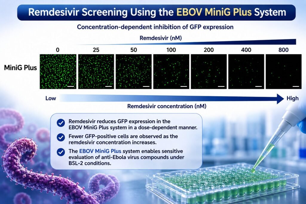
The HEK293T cells, after transfection with the EBOV MiniG Plus system, were treated with increasing drug concentrations and changes in the green fluorescent signals, produced by the GFP-positive cells, was measured
As found, Remdesivir produced concentration-dependent inhibition in both the conventional MiniG and MiniG Plus systems. Importantly, the enhanced MiniG Plus system produced a stronger initial fluorescent signal, making changes caused by antiviral treatment easier to detect.
The estimated EC50 values for remdesivir were similar between the two systems, approximately 87 nM for the conventional MiniG system and 84 nM for MiniG Plus.
In contrast, rupintrivir showed no inhibitory effect under the tested conditions, supporting the specificity of the assay.
A Better Platform for High-Throughput Screening
Z-factor, a statistical parameter and an indicator of the screening quality determined the suitability of the assay for high-throughput drug screening.
As found:
- The EBOV MiniG Plus system achieved a higher Z-factor of 0.72, compared with 0.52 for the conventional MiniG system, indicating improved assay performance and a greater ability to distinguish meaningful biological effects from experimental variability.
This improvement has practical implications for pharmaceutical research and antiviral drug discovery. A stronger and more reliable signal can improve the detection of candidate compounds, particularly during large-scale screening programs.
Practical Applications and Future Potential
EBOV MiniG Plus system is presented as a stronger and more sensitive platform for antiviral drug screening while maintaining its ability to measure the antiviral activity accurately.
- It provides a safer alternative for studying Ebola virus transcription and replication mechanisms under lower biosafety conditions.
- The improved GFP signal can increase the sensitivity and reliability of antiviral drug screening.
- The platform may reduce the cost and technical complexity associated with high-throughput screening.
- The concept may extend beyond Ebola virus research. Similar enhancement strategies could potentially be explored in minigenome systems developed for other high-risk RNA viruses.
As researchers continue to prepare for emerging infectious diseases, improved laboratory models such as EBOV MiniG Plus could help accelerate the discovery of new treatments while reducing dependence on experiments involving infectious high-risk viruses.