AI-Powered CAR-T Analysis Could Predict Cancer Immunotherapy Response
- Dr. Diksha Varma
- October 9, 2026
- 5 mins
Machine Learning and CAR-T Therapy: A New Approach to Predicting Cancer Immunotherapy Outcomes
CAR-T cell therapy has transformed cancer treatment by enabling a patient’s immune cells to recognize and attack malignant cells. However, an important challenge remains: how can researchers predict which CAR-T cells will work effectively before they are used clinically? Conventional approaches for evaluating CAR-T performance can require substantial time, resources, and laboratory effort, while their ability to predict clinical response can be limited.
Scientists from Rutgers University-New Jersey Medical School presents a machine learning (ML)-based approach that addresses these challenges by automatically measuring the quality of the CAR-T immunological synapse (IS). For this, high-resolution imaging, using a glass-supported planar lipid bilayer, was combined with artificial neural networks (ANNs) and multi-scale cell instance segmentation. This enabled automated analysis of thousands of CAR-T cell synapses rather than relying on manual evaluation of a limited number of cells.
The study further examined CAR-T cells obtained from patients who had received Kappa-CAR-T therapy. The ML-based analysis identified measurable differences in immunological synapse characteristics between a clinical responder and a non-responder, supporting the quality of CAR-T immunological synapse as a potential composite biomarker for evaluating CAR-T activity and provide a foundation for a proposed Synapse Predicts Efficacy (SPE) assay for research and future translational applications.
Machine Learning Could Improve CAR-T Cell Therapy Development
CAR-T therapy involves genetically modifying T cells so that they express chimeric antigen receptors capable of recognizing specific tumor-associated targets. Although this approach has produced significant clinical benefits in several blood cancers, CAR-T performance can vary substantially.
Researchers therefore need reliable ways to compare different CAR designs, identify promising cell products, and potentially determine which patients are more likely to respond. For this, researchers primarily focus on a feature that is directly related to CAR-T cell function: the immunological synapse. The immunological synapse is the organized interface formed when a CAR-T cell interacts with its target. Its structure, signaling, cytoskeletal organization, and distribution of cytotoxic molecules provide information about how effectively a CAR-T cell may respond to a tumor.
When a CAR-T cell encounters its target, multiple molecular and cellular processes occur at the contact site, including receptor engagement, F-actin accumulation, signaling through molecules such as phosphorylated zeta-chain protein (pZeta), tumor-antigen accumulation, and polarization of cytotoxic molecules such as perforin.
These features were captured by the researchers through high-resolution imaging and quantification was achieved using artificial intelligence.
Why Automating CAR-T Synapse Analysis Matters?
Till now, immunological synapse quality was assessed manually. However, manual image analysis is time-consuming, labour-intensive, and difficult to reproduce consistently. This conventional manual analysis typically evaluates fewer than 100 synapses, limiting the amount of information available for statistical analysis.
With the growing technological advancements in machine learning models, researchers found a faster alternative.
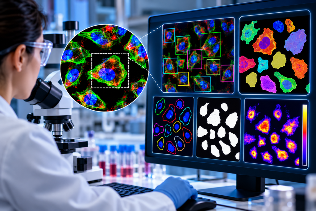
Instead of manually identifying and measuring every cell, the trained neural network:
- Detects individual cells
- Separates neighboring cells
- Generates segmentation masks
- Identifies contours
- Extracts quantitative fluorescence information.
This creates a scalable workflow capable of processing thousands of CAR-T immunological synapses.
Combining Microscopy, Biomarkers and AI for successful Cancer Immunotherapy Response
Kappa-CAR-T cells derived from patients who participated were used in the clinical trials. Two patient samples were selected as a proof-of-concept comparison: one was classified as a responder, whereas the other was classified as a non-responder. The study first characterized the cells using flow cytometry and examined CD4, CD8, CD3, CAR expression, and cell viability.
Then the CAR-T immunological synapse was recreated using a glass-supported planar lipid bilayer containing fluorescently labelled Kappa protein. This system provides a controlled surface that can mimic important aspects of the target-cell interaction while allowing researchers to obtain high-resolution images.
The CAR-T cells were labelled to visualize four major components:
- F-actin, which reflects cytoskeletal organization.
- Perforin, associated with cytotoxic activity.
- Tumor antigen, representing antigen accumulation at the synapse.
- pZeta, a key signaling component.
The resulting multi-channel confocal images provided a detailed representation of the CAR-T immunological synapse.
Artificial Neural Network-Based Image Analysis
The computational component consisted of two major stages: training and evaluation.
During training, manually segmented CAR-T images were used to train the artificial neural network. The researchers had 156 manually masked images and divided them into training, testing, and validation datasets.
The system incorporated object detection into instance segmentation to address difficult imaging conditions, including clustered cells, irregular cell shapes, cell overlap, low-contrast boundaries, and background noise.
During evaluation, the trained model generated:
- Bounding boxes
- Individual cell segmentation
- Cell contours
- Segmentation masks
- Fluorescence intensity measurements
The resulting masks were then applied across the different imaging channels to quantify immunological synapse characteristics.
The model used data augmentation, including cropping, flipping, random expansion, contrast distortion, and brightness distortion. The network was trained using the Adam optimizer for up to 100 epochs.
Testing the accuracy of the Machine Learning Model
Referred to as InstSeg, researchers compared their instance-segmentation approach with other approaches, including DCAN, CosineEmbedding, and Mask R-CNN, showcasing that InstSeg performed strongly across the evaluated conditions.
For example, with 75% of the training data, InstSeg achieved an AP@0.7 of 61.65% for bounding-box evaluation, compared with 30.87% for DCAN and 43.94% for Mask R-CNN.
For segmentation evaluation using 75% of the data, InstSeg achieved an AP@0.7 of 63.03%, compared with 24.71% for DCAN and 57.69% for Mask R-CNN.
The researchers reported that InstSeg had an average accuracy advantage of 2.86% over Mask R-CNN across the evaluated scenarios.
These results demonstrate the value of combining object detection and instance segmentation for automated CAR-T cell image analysis.
Evaluation of ML trained Model on the Patient Samples
The researchers applied the trained model to CAR-T cells obtained from the two patients.
The system detected 2,127 cells from one patient and 2,404 cells from other, providing a substantially larger dataset for comparison than would typically be practical with manual analysis.
The analysis showed significant differences in several immunological synapse measurements:
- For the perforin channel, the total intensity was 647,860.7 ± 15,339.5 for responders compared with 375,259.0 ± 7,196.0 for non-responders.
- For the tumor-antigen channel, the corresponding values were 579,608.5 ± 14,964.7 and 478,515.0 ± 10,001.9.
- For pZeta, the values were 1,008,681.9 ± 32,211.0 and 706,055.1 ± 22,955.2, respectively.
The study reported statistically significant differences between the two patients for these channels and the results were found to be consistent even at different tumor-antigen concentrations.

Applications of the AI-Powered CAR-T Technology
The most important practical application is the possibility of developing a rapid, automated CAR-T efficacy assessment platform.
The proposed SPE assay could potentially help researchers compare CAR constructs before investing substantial resources in more complex preclinical or clinical studies.
- CAR-T construct optimization
Researchers could use immunological synapse quality as an additional quantitative parameter when comparing different CAR designs, including changes to CAR architecture and signaling components.
- Preclinical CAR-T screening
Instead of relying exclusively on lengthy killing assays or animal models, researchers could potentially use automated synapse measurements as an early-stage screening tool.
The study specifically proposes that the approach could help guide the design and optimization of CAR constructs for potential clinical development.
- High-throughput image analysis
Automated segmentation allows thousands of cells to be analyzed, creating larger datasets than manual analysis can typically provide.
- Potential biomarker development
The findings support CAR-T immunological synapse quality as a potential composite biomarker associated with antitumor activity.
However, the study presents this as a promising research direction rather than an established clinical diagnostic test.
- Translational immunotherapy research
The same computational framework could potentially be adapted for research involving other CAR-modified immune cells and immune-cell therapies, specifically, the CAR-T and CAR-NK applications and the potential relevance to other immune products.
Future Insights
The study successfully demonstrates how artificial intelligence, quantitative microscopy, and immunology can be integrated into a single analytical workflow.
Though the presented technology is not yet a clinically validated predictor of CAR-T treatment response but its ability to automate image analysis, increase measurement throughput, and provide quantitative information about CAR-T cell function could make it useful for CAR design, preclinical screening, immunotherapy research, and future biomarker development.
However, some limitations could be addressed in future, in the direction of the existing outcomes.
- Evaluations with the larger sample sizes will strongly demonstrate the application of machine learning-based quality of the CAR-T immunological synapse (IS).
- The system can incorporate a complete tumor-cell environment with other co-stimulatory and co-inhibitory molecules to assess the assess the effects of intratumor heterogeneity on immunological synapse quality.
- The SPE method needs to be compared with conventional CAR-T evaluation approaches.
The study, thus, provides a proof-of-concept for automated CAR-T synapse analysis, providing a compelling demonstration of how machine learning can improve the quantitative analysis of CAR-T immunological synapses.