Artificial Intelligence Enhances Breast Cancer diagnosis through Deep Learning
Why Traditional Breast Cancer Diagnosis Needs Improvement
Breast cancer remains one of the leading causes of cancer-related deaths among women worldwide. Early and accurate diagnosis plays a critical role in improving survival rates and guiding effective treatment. However, traditional histopathological grading where pathologists examine tissue samples under a microscope that can be subjective, leading to inconsistencies between observers.
Histological grading is one of the most important factors in determining the aggressiveness of breast cancer. Pathologists evaluate tissue samples stained with hematoxylin and eosin (H&E) using the Nottingham Histological Grading System.
While this approach is well established, it has several limitations:
- Variability between different pathologists
- Inconsistent grading by the same observer over time
- Time-intensive manual image analysis
- Dependence on expert availability
These challenges can influence treatment decisions and patient outcomes.
How Deep Learning and CNNs is improving diagnostic accuracy
A recent study conducted by scientists of Princess Nourah bint Abdulrahman University, Riyadh, explores how deep learning for breast cancer detection can address these challenges.
The researchers developed an artificial intelligence (AI) system based on Convolutional Neural Networks (CNNs) to automate the histological classification of breast cancer using medical images. The application of AI could improve diagnostic consistency, reduce manual workload, and potentially make high-quality breast cancer screening more accessible, especially in underserved regions.
Rather than replacing pathologists, the proposed system is designed to function as a clinical decision-support tool by:
- Identifying high-quality tissue regions
- Eliminating low-quality images
- Automatically classifying breast cancer tissue
- Reducing subjective interpretation
How Artificial Intelligence (AI) system detects Breast Cancer
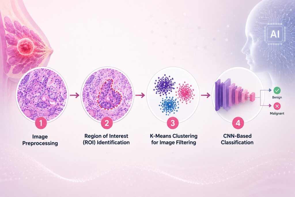
A multi-stage image processing and deep learning workflow combines traditional image analysis and modern AI techniques.
- Image Preprocessing
The first step involved preparing histopathological images for analysis.
Since medical images often contain artifacts, blurred regions, or insufficient tumor tissue, preprocessing is essential to improve model performance.
The preprocessing pipeline include:
- Image normalization
- Color standardization
- Noise reduction
- Separation of hematoxylin (H) and eosin (E) staining channels
By isolating the hematoxylin channel, which highlights cell nuclei, the model focus on the most informative cellular structures associated with breast cancer.
- Region of Interest (ROI) Identification
Not every part of a pathology slide contains useful diagnostic information.
Image segmentation techniques identify Regions of Interest (ROIs), ensuring that CNN analyzed tissue areas are rich in tumor cells while ignoring background regions and irrelevant artifacts. This significantly improves data quality before model training.
- K-Means Clustering for Image Filtering
An innovative aspect of the AI system was the use of the K-means clustering algorithm.
Instead of feeding every image into the neural network, K-means classifies pixels based on similarity to create segmentation masks. Images containing insufficient tumor cells or poor-quality tissue are filtered out automatically.
This preprocessing step ensures that only informative images are used for training, improving both computational efficiency and classification accuracy.
- CNN-Based Classification
After preprocessing and filtering, the remaining high-quality images are used to train a Convolutional Neural Network.
CNNs are particularly effective in medical image analysis because they automatically learn visual features without requiring handcrafted feature extraction. The network learn to recognize patterns associated with different breast cancer histological grades, enabling automated classification.
Outcomes of application of AI systems
Improved Classification Performance
Compared with AlexNet, VGG16, GoogLeNet, and LeNet, the AI system model achieves:
- Top-5 Accuracy: 5%
- Top-1 Accuracy: 0%
These results indicate stronger classification performance while maintaining a relatively efficient network architecture.
Better Image Quality Control
One of the most valuable outcomes are the ability to automatically distinguish useful histopathological images from poor-quality samples. Filtering non-informative images before CNN training reduces noise in the dataset and improves model learning.
Enhanced Diagnostic Consistency
As AI evaluates every image using the same computational rules, the system minimizes variability introduced by human interpretation. This could improve consistency across laboratories and support standardized pathology workflows.
Efficient Feature Extraction
Separating hematoxylin and eosin staining enables the CNN to focus primarily on nuclear features, which are highly relevant in cancer grading. This targeted feature extraction improves the model’s ability to identify meaningful pathological patterns.
Implications of Deep Learning & CNNs across healthcare system
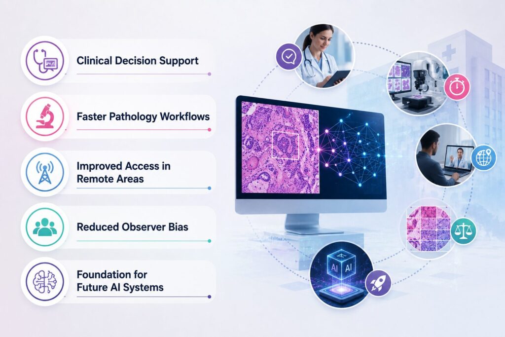
Clinical Decision Support: The AI system can serve as a second reader, highlighting suspicious regions and assisting specialists in making faster, more consistent diagnoses.
Faster Pathology Workflows: Automated image filtering reduces the time spent reviewing unusable or poor-quality images, allowing clinicians to focus on diagnostically relevant cases.
Improved Access in Remote Areas: The AI system could be developed into a low-cost, portable solution for healthcare professionals working in rural or underserved regions where pathology expertise is limited.
Reduced Observer Bias: Since the CNN applies standardized classification criteria, it helps reduce the subjective variability commonly associated with manual histological grading.
Foundation for Future AI Systems: The methodology can also be adapted for diagnosing other cancers and diseases using histopathological images, making it a scalable approach for digital pathology.
Future Insights
As digital pathology continues to evolve, intelligent systems like this have the potential to support clinicians, accelerate diagnoses, and ultimately improve patient outcomes through more accurate and reproducible breast cancer assessment.
While the results are promising, several areas for improvement could be focussed:
- Validating the model using larger and more diverse datasets.
- Investigation of additional deep learning architectures, such as deeper neural networks and hybrid AI models.
- Development of more automated image preprocessing workflows to reduce manual intervention.
- Exploring alternative image normalization and segmentation techniques.
- Translation of the research into user-friendly clinical software suitable for routine healthcare settings.